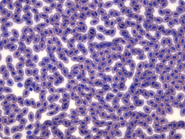
Normal Fish Blood Smear Stained with Diff-Quik
A bright field light micrograph of a normal blood smear from a rainbow trout, stained using Diff-Quik. The image highlights the typical appearance of healthy fish blood cells, including erythrocytes and leukocytes, serving as a reference for identifying abnormalities during disease diagnostics.
Host: Rainbow Trout
Host Tissue: Blood
Photographer / Illustrator: Stephen Atkinson
Image Type: Light Micrograph (Bright Field, Diff-Quik)